METASTATIC RENAL CELL ADENOCARCINOMA OF THE GALLBLADDER: A CASE REPORT
Abstract
Background: Gallbladder (GB) involvement in patients with renal cell carcinoma (RCC) is extremely rare.
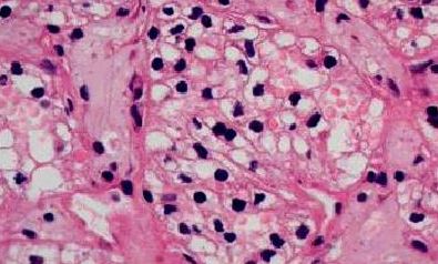
Case report: We present a report of a 58-year-old man with a metachronous metastatic contralateral renal cell adenocarcinoma of the gallbladder. The patient presented with the acute right upper quadrant pain and cholecystitis three years after resection of the primary renal neoplasm. Gallbladder tumor was detected clinically. The tumor was located in the fundus of the gallblladder under the lamina propria mucosa with subserosal invasion. Blood biochemistry results were in normal ranges as well as carcinoembryonic antigen (CEA) and carbohydrate antigen 19-9 (Ca 19-9). A histopathological examination revealed that the mucosal surface was covered with yellowish fibronecrotic tissue but contained no tumor cell. The tumor was located under the lamina propria mucosa with subserosal invasion. Tumor cells were positive for PAS, vimentin and CD 10, but negative for carcinoembryonic antigen (CEA), carbohydrate antigen (Ca 19-9) and cytokeratin 7 (CK7). Additional specific and immunohistochemical tests confirmed that gallbladder tumor was a metastasis of renal cell carcinoma. Renal cell adenocarcinoma was specified as Fuhrman's grade 2.
Conclusions: Pre-operative imaging studies are often futile for the differentiation between primary and secondary tumors of the gallbladder. Primary tumors of the gallbladder often coexist with gallstones while polypoid lesions in an acalculous gallbladder are frequently associated with metastasis.
Keywords
DOI: 10.5457/ams.v38i1.39
